|
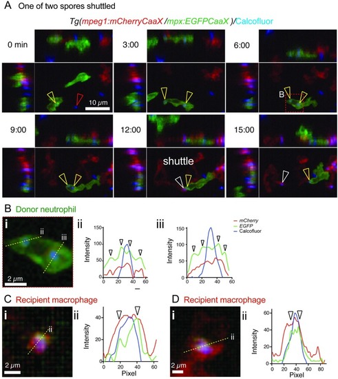
Shuttling of <italic>T</italic>. <italic>marneffei</italic> conidia between neutrophils and macrophages involves phagosome transfer.(A) Shuttle of calcofluor-stained conidium (blue) from Tg(mpx:EGFP-CaaX) neutrophils (green) to Tg(mpeg1:mCherry-CaaX) macrophages (red). These reporter lines have membrane-localized fluorophore expression. Panels include isometric orthogonal yz and xz views corresponding to the xy maximal intensity projection and indicate the time in min from start of the movie. Colored arrowheads indicate a conidium before it is phagocytosed by the donor neutrophil (red), conidia within the donor neutrophil (yellow), and the conidium at the point of intercellular transfer and within the recipient macrophage (white). (Bi) Detail of the boxed area of the donor neutrophil in (A), 6-min panel. Yellow dotted line indicates the position of the cross-section for the 3-color fluorescence intensity plots in (ii) and (iii). Both shuttled and nonshuttled conidia are flanked by peaks of green fluorescence, consistent with their location in a membrane-lined phagosome. (C,D) Cross-sections fluorescence intensity profiles (ii) corresponding to the yellow lines in (i) for 2 macrophages that received a spore from a neutrophil in this dataset, which contained 3 independent spore shuttles. The arrowed EGFP-channel signal demonstrates the transfer of neutrophil-derived EGFP-tagged membrane in the vicinity of the spore (blue channel signal). Scales as shown. Stills in A correspond to S3A Movie. EGFP, enhanced green fluorescent protein; mpeg1, macrophage-expressed gene 1; mpx, myeloid-specific peroxidase; Tg, transgenic.
|