|
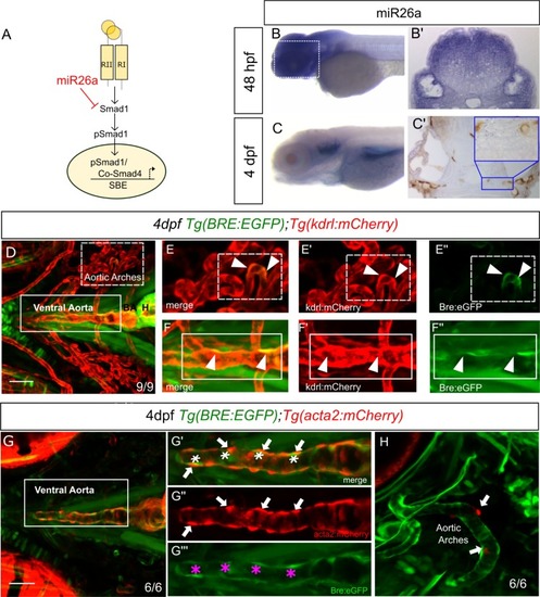
<italic>miR26a</italic> is expressed in blood vessels; endothelial cells have active BMP signalling.A) Model of how miR26a controls BMP signaling via direct targeting of smad1. B) Lateral view of whole mount in situ expression of miR26a at 48 hpf shows ubiquitous expression pattern, with strong expression in the ventral head of the embryo. B’) Cross section of the head at 48 hpf. C) At 4 dpf miR26a is expressed in the pharyngeal arches, bulbous arteriosus and ventral aorta. C’) Cross section of the head showing miR26a expression in blood vessels (purple; punctate stain) compared with endothelial stain (brown; kdrl:GFP transgenic). Inset is an enlargement of image in C’. D) Ventral view of the pharyngeal region of a 4 dpf double transgenic Tg(BRE:EGFP);Tg(kdrl:mCherry) embryo shows BRE:EGFP (green) expression within endothelial cells in aortic arches (red, white arrowheads in E’-E”‘) and ventral aorta (red, white arrowheads F’-F”‘). G-H) Ventral and lateral views of a 4 dpf double transgenic Tg(BRE:EGFP); Tg(acta2:mCherry) zebrafish shows that acta2 positive cells are in direct contact with BMP-responsive endothelial cells but do not express BRE:EGFP. Scale bar represents 50μm.
|