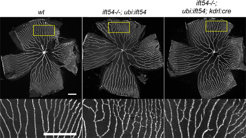
Endothelial‐specific excision of the Tg(ubi:loxP‐ift54‐loxP‐myr‐mcherry,myl7:EGFP)sh488‐rescuing transgene causes no apparent further impairment to adult retina vasculature in ift54tp49; Tg(ubi:loxP‐ift54‐loxP‐myr‐mcherry,myl7:EGFP)sh488 fish. Confocal stack projections showing endothelial EGFP expression in flat‐mount views of retina from sibling four‐month‐old Tg(ubi:loxP‐ift54‐loxP‐myr‐mcherry,myl7:EGFP)sh488 /+;Tg(fli1a:EGFP)y1 /+ (n = 5) (wt) and ift54tp49; Tg(ubi:loxP‐ift54‐loxP‐myr‐mcherry,myl7:EGFP)sh488 /+; Tg(fli1a:EGFP)y1 /+ (n = 4) (ift54−/−;ubi:ift54) and ift54 tp49; Tg(ubi:loxP‐ift54‐loxP‐myr‐mcherry,myl7:EGFP)sh488 /+;Tg(kdrl:cre)s898/+; Tg(fli1a:EGFP)y1 /+ (n = 5) (ift54−/−;ubi:ift54;kdrl:cre) fish. The yellow boxes indicate the region of retina enlarged below. The Tg(kdrl:cre)s898 /+ caused no apparent further impairment to the vasculature in the fish with partial transgenic rescue of ift54 tp49. EGFP, enhanced green fluorescent protein; WT, wild‐type. Scale bars = 300 μm
|
