
Fig. 2
- ID
- ZDB-FIG-190809-21
- Publication
- Pitchai et al., 2019 - Zebrafish as an Emerging Model for Bioassay-Guided Natural Product Drug Discovery for Neurological Disorders
- Other Figures
- All Figure Page
- Back to All Figure Page
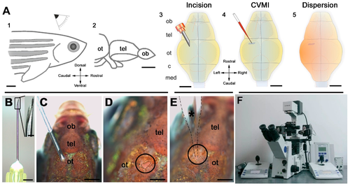
Outline of the pattern and its target regions of cerebroventricular microinjection (CVMI). (A) CVMI is achieved at the dorsal surface of the head (1) and targets, in this example, the forebrain that is rostral to the optic tectum (2). For injection, an incision is made into the skull over the optic tectum using a barbed-end canula (3). Through this slit, liquid is injected using a glass capillary (4). Injected liquid disperses rostrally (5). (B) The canula used for incision. (C) The incision on an adult fish (dorsal view). (D) The incision site marked by dashed lines. (E) Injection with the glass capillary (*) (dotted lines mark the outline). (F) Injection apparatus. Images (A–E) are adapted from [132]. (c: cerebellum; med: medulla; ob: olfactory bulb; ot: optic tectum; and tel: telencephalon) |