
Fig. 1
- ID
- ZDB-FIG-190806-18
- Publication
- Baumgartner et al., 2019 - Identification of regulatory elements recapitulating early expression of L-plastin in the zebrafish enveloping layer and embryonic periderm
- Other Figures
- All Figure Page
- Back to All Figure Page
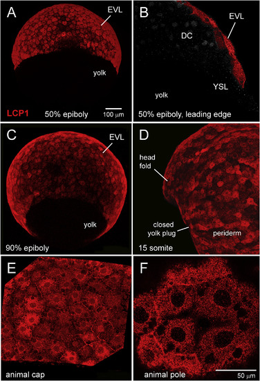
Immunohistochemical survey of L-plastin expression in early zebrafish development. A. Enveloping layer (EVL) expression at 50% epiboly, seen in equatorial view. B. EVL expression at 50% epiboly, seen in cutaway view of a single focal plane. The squamous EVL is strongly stained, in contrast to the deep cells (DC) and the yolk syncytial layer (YSL). C. EVL expression at 90% epiboly, seen obliquely from the vegetal pole. D. Periderm expression at the somite stage. Stain intensity is variegated, appearing brighter or darker in adjacent cells. E. 70% epiboly, intermediate magnification of animal cap EVL. F. 70% epiboly, high magnification of animal cap EVL. In this single focal plane, L-plastin expression appears as tightly packed cytoplasmic dots or clumps. DC = deep cells; EVL = enveloping layer; YSL = yolk syncytial layer. |
Reprinted from Gene expression patterns : GEP, 32, Baumgartner, E.A., Compton, Z.J., Evans, S., Topczewski, J., LeClair, E.E., Identification of regulatory elements recapitulating early expression of L-plastin in the zebrafish enveloping layer and embryonic periderm, 53-66, Copyright (2019) with permission from Elsevier. Full text @ Gene Expr. Patterns