
Fig. S2
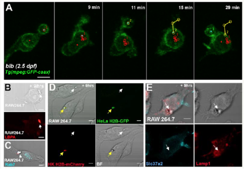
Molecular characterization of the gastrosome and its enlargement in slc37a2 mutant embryos- Related to Figure 4 and Figure 5 (A; related to Fig.4 F and movie 5) SPIM time-lapse of 2.5 dpf blbNY007 microglia labeled with Tg(mpeg:GFP-caax). The red dot marks the growing vesicle. Red, yellow and blue tracks show the trajectories of three incoming phagosomes. Time indicated in minutes. Scale bar 10μm. (B) Fixed RAW264.7 macrophages, 9hrs after apoptosis induction in unlabeled HK and HeLa cells. Immunostaining against LBPA (red). The white arrow indicates the collection compartment (gastrosome). Scale bars 5μm. (C) Fixed RAW264.7 macrophages, 9hrs after apoptosisb induction in unlabeled HK and HeLa cells. Immunostaining against Rab7 (cyan). The white arrow indicates the collection compartment (gastrosome). Scale bars 5μm. (D) RAW264.7 macrophages fixed 9 hours after apoptosis induction of nuclear-labelled HK H2B-mCherry (red) and nuclear labeled HeLa H2B-GFP (green). The image shows two cells with a collection compartment (gastrosome) each. In one the cell gastrosome is fluorescently labelled (yellow arrow) while the other is not (white arrow) as it contains cellular material that is not fluorescently labelled. Scale bars 5μm. (E) RAW264.7 macrophages fixed 9 hours after apoptosis induction of unlabelled HeLa cells. Immunostaining of Lamp1 (red) and Slc37a2 (cyan). The white arrow points to a collection compartment (gastrosome) that is Lamp1 positive and Slc37a2 negative. |
Reprinted from Developmental Cell, 49(1), Villani, A., Benjaminsen, J., Moritz, C., Henke, K., Hartmann, J., Norlin, N., Richter, K., Schieber, N.L., Franke, T., Schwab, Y., Peri, F., Clearance by Microglia Depends on Packaging of Phagosomes into a Unique Cellular Compartment, 77-88.e7, Copyright (2019) with permission from Elsevier. Full text @ Dev. Cell