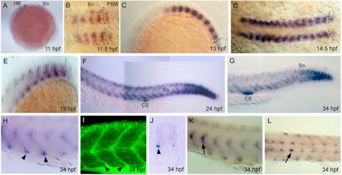
Expression of uncx4.1 gene during somitogenesis. Whole-mount in situ hybridization of uncx4.1 at (A) 11 hours post fertilization (hpf), (B) 11.5 hpf, (C) 13 hpf, (D) 14.5 hpf, (E) 19 hpf, (F) 24 hpf, and (G–L) 34 hpf. (A, C, E–H, J, K) Lateral view, (B, D) dorsal view, and (I) transversal section. (A–H, J–L) Anterior to left, and (I) toward viewer. (A) Expression in early somites (So) at 11 hpf. HB = hindbrain. (B–D) Expression in somites from 11.5 to 14.5 hpf. Dotted lines show boundary between presomitic mesoderm (PSM) and last formed somite. (E) Expression is lost in the myoseptum and is restricted posteriorly in developing somites at 10 hpf. (F) Expression is restricted to dorsal and ventral margins of anterior somites at 24 hpf, and (G) disappears dorsally at 34 hpf. CS = Corpuscle of Stannius, So = somites. (H) Arrowheads indicate in situ hybridization staining in ventro-latero-posterior cells (VLP) at 34 hpf. (I) Arrowheads indicate VLP cells in phalloidin-stained somites at 34 hpf. (J) Arrowhead indicates expression in VLP cells in transversal section at 16th somite level at 34 hpf. (K, L) Arrows indicate expression in sclerotome cells in (K) lateral and (L) dorsal view at 34 hpf.
|
