FIGURE

Figure 3

ID
ZDB-FIG-190723-485
Publication
Li et al., 2019 - Methylglyoxal-Induced Retinal Angiogenesis in Zebrafish Embryo: A Potential Animal Model of Neovascular Retinopathy
Other Figures
All Figure Page
Back to All Figure Page
Figure 3

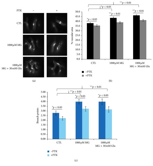
Effect of VEGF receptor inhibitor (PTK787) in MG-induced zebrafish. (a) Fluorescence microscope observations shown added 0.5 µM PTK787 in 1 dpf in the 1000 µM MG and 30 mM glucose treatment group reverted blood vessel hyperbranching in zebrafish embryos at 4 dpf. The proangiogenic effect induced by MG treatment was inhibited and exhibited no significant difference compared with the no-treatment control. Quantification of the percentage of vascular area (b) and number of branch points (c) in midperiphral retina by PTK787, MG, and glucose treatment in zebrafish embryos at 4 dpf. n = 30 eyes of 15 zebrafish embryos per group; mean ± SEM. p  <  0.05, ∗∗p < 0.01 vs control group. p > 0.05 nonsignificant (NS). Data were analyzed using the one-way ANOVA and then Student's t-test. Glu, glucose. CTL, control.

Expression Data

Expression Detail
Antibody Labeling
Phenotype Data
Fish:
Condition:
Observed In:
Stage: Day 4

Phenotype Detail
Acknowledgments
This image is the copyrighted work of the attributed author or publisher, and ZFIN has permission only to display this image to its users. Additional permissions should be obtained from the applicable author or publisher of the image. Full text @ J Ophthalmol