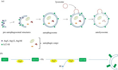
(a) Autophagosome formation. Schematic of autophagosome formation and degradation: Within the cytoplasm, double-membraned, sac-like structures called phagophores are the first morphologically recognizable autophagic precursors and can be distinguished within cells by the proteins that associate with their membranes. A complex comprising ATG12–ATG5–ATG16L1 proteins enables the conjugation of LC3-II to the membranes. The edges of the phagophore elongate and eventually fuse while engulfing a portion of the cytoplasm. As the phagophore enlarges and approaches closure, the ATG5–ATG12–ATG16L1 complex dissociates from the outer membrane, whereas LC3-II remains associated. The resulting structure is a spherical double-membrane organelle, called the autophagosome. Following closure, autophagosomes are trafficked by dynein motors along microtubules to the perinuclear region where they fuse with the lysosomes and their contents are degraded. (b) Lipidation of LC3-II. During autophagosome formation, LC3 (and other ATG8 ubiquitin-like family proteins) are conjugated to the lipid PE in autophagosome membranes. This lipidation requires a protease and two ubiquitin-like conjugation systems (explained in detail in [1,2]). ATG4 is a cysteine protease which cleaves the C-terminus of LC3 exposing a glycine residue. This first cleaved form of LC3 is called LC3-I. A further reaction then occurs involving a complex of ATG12–5 and ATG16L1, which together act as an E3-like ligase. This determines the site of LC3 lipidation and assists the transfer of LC3-I to PE in membranes to form LC3-II. ATG8/LC3 proteins may assist in the expansion and closure of autophagosomal membranes, in autophagosome-lysosome fusion and inner autophagosomal membrane degradation.
|
