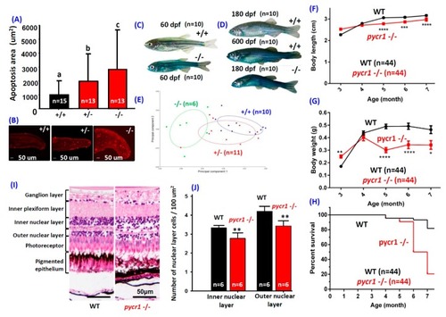
Morphologies of pycr1 gene knockout (KO) Zebrafish. Quantitative (A) and qualitative (B) detection of fluorescence apoptotic intensity within tail in the pycr1 KO fish embryos aged at 24 hour-post-fertilization (hpf) by Terminal deoxynucleotidyl transferase dUTP nick end labeling (TUNEL) staining. Data were presented with mean ± SEM and the significance was tested by one-way ANOVA; n = 13–15. The label above column with different letter means reaching significant difference with p < 0.05. (C) The detection of intestinal integrity in the pycr1 KO fish at 60 day-post-fertilization (dpf) by Smurf dye staining. (D) Detection of intestinal integrity in the WT and pycr1 KO fish at 180 dpf or natural aging WT fish aged at 600 dpf by Smurf dye staining. (E) The Principle Component Analysis (PCA) plot for morphometric analyses among the WT (+/+), heterozygotic (+/−), and homozygotic (−/−) pycr1 KO fish aged at 180 dpf. (F) The growth curves in body length of the WT and the pycr1 KO fish. (G) The growth curves in body weight of the WT and the pycr1 KO fish. (H) The mortality curves of WT and pycr1 KO fish. (I) The retina histological comparison between WT and pycr1 KO fish aged at 180 dpf. Data were presented with mean ± SEM and the significance was tested by t-test in (F–H), n = 44; * p < 0.05, ** p < 0.01, *** p < 0.005, and **** p < 0.001. (J) The quantitative comparison of retina cell density between WT and pycr1 KO fish. The data were presented with mean ± SEM and the significance was tested by t-test, n = 6; ** p < 0.01.
|
