FIGURE

Figure 3

ID
ZDB-FIG-190723-2407
Publication
De La Garza et al., 2017 - Concise Review: Hematopoietic Stem Cell Origins: Lessons from Embryogenesis for Improving Regenerative Medicine
Other Figures
All Figure Page
Back to All Figure Page
Figure 3

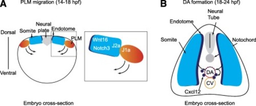
Somitic signals regulate hematopoietic stem cell (HSC) formation in zebrafish. (A): Cross‐sectional view of a zebrafish embryo at 14 hpf. The posterior lateral mesoderm migrates medially, sliding under the somites, where it receives Wnt16 and Notch3 signals that promote hematopoietic induction. The close proximity of PLM and somite cells is achieved by heterotypic interactions between Jam1a on PLM and Jam2a on somites, which helps to strengthen the Notch signal. (B): Cross‐sectional view of a zebrafish embryo at 24 hpf. Anterior portions of somites give rise to the endotome, which contributes cells to the dorsal aorta that promote HSC formation, in part by Cxcl12 production. Curved arrows denote the movement of cells. Abbreviations: CV, caudal vein; DA, dorsal aorta; hpf, hours postfertilization; J1a, Jam1a; J2a, Jam2a; PLM, posterior lateral mesoderm.

Expression Data

Expression Detail
Antibody Labeling
Phenotype Data

Phenotype Detail
Acknowledgments
This image is the copyrighted work of the attributed author or publisher, and ZFIN has permission only to display this image to its users. Additional permissions should be obtained from the applicable author or publisher of the image. Full text @ Stem Cells Transl. Med.