FIGURE

Fig. 1

ID
ZDB-FIG-190723-2397
Publication
Corallo et al., 2016 - The zebrafish as a model for studying neuroblastoma
Other Figures
All Figure Page
Back to All Figure Page
Fig. 1

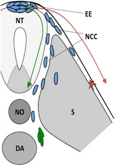
Patterns of NCC migration in zebrafish. Schematic cartoon depicting the two main migratory pathways of NCCs during embryonic development (transverse section of a vertebrate embryo). NCCs (blue) follow the ventromedial pathway (green arrow) and migrate between the neural tube and somites and then bilaterally reach the dorsal portion of the dorsal aorta, where they differentiate into sympathoadrenal progenitors (green). NCCs that migrate dorsolaterally (red arrow) between somites and the overlying ectodermal epithelium give rise to pigment cells (red). NCCs neural crest cells, EE ectodermal epithelium, NT neural tube, NO notochord, DA dorsal aorta, S somite

Expression Data

Expression Detail
Antibody Labeling
Phenotype Data

Phenotype Detail
Acknowledgments
This image is the copyrighted work of the attributed author or publisher, and ZFIN has permission only to display this image to its users. Additional permissions should be obtained from the applicable author or publisher of the image. Full text @ Cancer Cell Int.