FIGURE

Figure 5

ID
ZDB-FIG-190723-2338
Publication
Gramage et al., 2014 - The expression and function of midkine in the vertebrate retina
Other Figures
All Figure Page
Back to All Figure Page
Figure 5

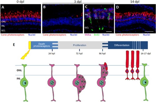
Midkine expression in zebrafish retinal regeneration. A: Cone photoreceptors are immunolabelled (red signal) in an unlesioned retina. B: Photoreceptors are nearly completely killed following exposure to a photolytic lesion. C: In situ hybridization showing that mdka is expressed in proliferating photoreceptor progenitors (pink label) following photoreceptor death. The mdka message co-localizes with antibody staining for bromodeoxyuridine (BrdU, green signal). D: Fourteen days following the onset of a photolytic lesion, rod (not shown) and cone photoreceptors (red signal) are regenerated. E: Timeline of the different processes that occur in the adult zebrafish retina after light-induced damage and the proliferative response of Müller glia. The pink colour represents the onset and duration of mdka expression in Müller glia and photoreceptor progenitors. Scale bar = 25 μm (C) and 50μm (A, B and D). GCL, ganglion cell layer; INL, inner nuclear layer; ONL, outer nuclear layer; dpl: days post-lesion.

Expression Data

Expression Detail
Antibody Labeling
Phenotype Data

Phenotype Detail
Acknowledgments
This image is the copyrighted work of the attributed author or publisher, and ZFIN has permission only to display this image to its users. Additional permissions should be obtained from the applicable author or publisher of the image. Full text @ Br. J. Pharmacol.