|
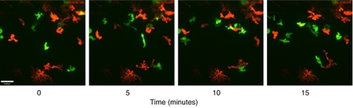
In vivo imaging of zebrafish immunity. Neutrophils (green) and macrophages (red) can be visualised during inflammation in vivo, showing clear differences in cell morphology and behaviour. Dynamic interaction between these cell types can also be visualised as they participate in the inflammatory process. Time-course images show a day-3 Tg(mpx:GFP)i114;Tg(fms:gal4)i186;Tg(UAS:nfsB.mCherry)i149 larva. A single immotile xanthophore can be seen at the bottom of the image. Images were acquired on a Perkin Elmer UltraVOX spinning disc confocal mounted on an Olympus IX81 microscope using a 40× oil immersion lens, NA 1.3. The movie from which these stills were acquired is available as supplementary material Movie 1, and a 3D reconstruction of the data is available as supplementary material Movie 2. Scale bar: 25 μm.
|