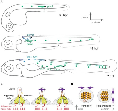
Development and organization of the posterior lateral-line sensory receptors. (A) Lateral view of a developing zebrafish at around 30, 48 hpf and 7 dpf (days post-fertilization) showing the development of the mechanoreceptive neuromasts that form the posterior lateral line. primI, first primordium; primII, second primordium; primD, dorsal primordium. (>) and (∧) indicate parallel and perpendicular neuromasts, respectively. (B) Lateral view of a neuromast. The hair-bundle comprises the kinocilium (K) and the stereocilia (S), and is contained within a gelatinous cupula. A neuromast contains two populations of hair cells of opposing hair-bundle polarities. Thus, a water movement (blue arrow) bending the cupula in a given direction depolarizes (+) one population of hair cells, whereas hyperpolarizes (-) the other one. This is translated into an increase or a decrease of the firing rate of the afferent neuron associated to each hair-cell population. (C) Top view of a parallel and a perpendicular neuromast, which are sensitive to water movements across orthogonal axes.
|
