|
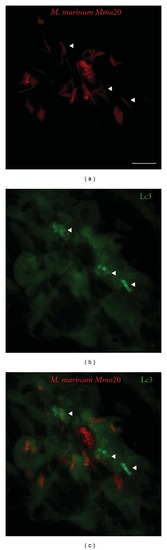
In situ detection of autophagy by Lc3 accumulation. CMV::LC3-GFP transgenic [15] zebrafish embryos (28 hpf) were injected into the caudal vein with 200 colony-forming units (CFU) of M. marinum Mma20 expressing a pMST3::mCherry vector. Confocal images were taken of a tail region of the developing larva at 3 days after infection (3 dpi), a point at which the M. marinum infection (a) has been established. Low levels of Lc3-GFP signal (b) can be observed throughout the cells, whilst brighter regions (indicated by arrowheads) are only observed upon Lc3 accumulation and formation of autophagic membranes associated with bacteria (c). Scale bar: 10 μm.
|