FIGURE

Fig. 2

ID
ZDB-FIG-190723-2033
Publication
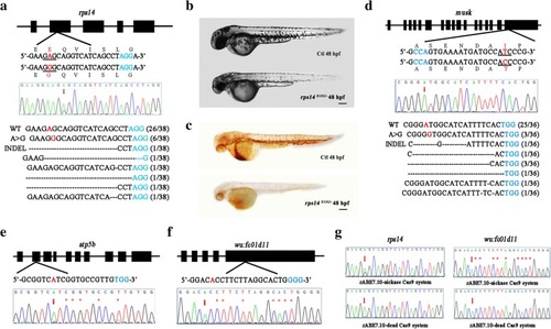
Qin et al., 2018 - Precise A•T to G•C base editing in the zebrafish genome
Other Figures
All Figure Page
Back to All Figure Page
Fig. 2

Codon-optimized zABE7.10 system in zebrafish induces A to G base conversion in zebrafish. a Schematic view of the gRNA target site in rps14 gene and the sequencing results. b Morphological phenotype of rps14E12G/− deficient embryos. co-dianisidine staining results of the rps14E12G/− embryos. d Schematic view of the gRNA target site in musk gene and the sequencing results. e Sequence chromatograms of atp5b locus. f Sequence chromatograms of wu:fc01d11 locus. g Comparison the base editing efficiency between zABE7.10-nickcase Cas9 and zABE7.10-dCas9 system. Target sequence (black), PAM region (blue), and the substituted bases (red) are indicated. The black line presents the changed amino acid. The red arrows indicate the overlapped peaks. The black dashes and blue dashes denote deleted bases and inserted bases in the sequence, respectively. The red asterisks indicated the overlapping peaks outside the targeted A

Expression Data

Expression Detail
Antibody Labeling
Phenotype Data

Phenotype Detail
Acknowledgments
This image is the copyrighted work of the attributed author or publisher, and ZFIN has permission only to display this image to its users. Additional permissions should be obtained from the applicable author or publisher of the image. Full text @ BMC Biol.