|
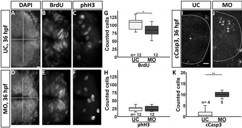
fgf3 impairment results in reduced proliferation and increased cell death in the posterior hypothalamus at 36 hpf. (A–F,I,J) Confocal maximum intensity projections of uninjected control (UC) and fgf3 morphant (MO) siblings immunostained for BrdU and phospho-histone H3 (phH3) counterstained with DAPI, or for cleaved caspase 3 (cCasp3) at 36 hpf. Dashed lines indicate ventricle in A and D, and outer posterior border of the hypothalamus in I and J. Examples of cCasp3 positive cells are indicated (arrowheads). Anterior is to the left. Scale bars: 10 µm. (G,H,K) Quantifications of BrdU, phH3 and cCasp3 positive cells after fgf3 impairment. Tukey boxplots show median, 25–75% percentile, IQR whiskers and outliers. n=number of analysed individuals. *P>0.05, **P>0.01.
|