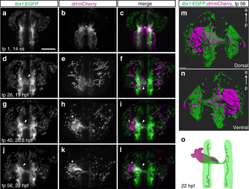
A tbx1+ sheath forms at the base of the FHF-derived heart tube. a–l Maximum intensity projections of representative stages from SPIM-imaged tbx1:EGFP;drl:mCherry double-positive transgenic embryos; dorsal views, anterior to the top. Imaging was initiated at 14 ss (16 hpf) and cardiac development followed until linear heart tube (LHT) stage (22–23 hpf, n = 3). a–c 14 ss stage embryo at the onset of medial FHF migration (asterisk). d–f The forming cardiac disk already contains tbx1:EGFP-positive cells (arrowhead). g–ltbx1:EGFP-positive cells (arrowhead in g, i) assemble at the base of the extending drl reporter-expressing heart cone (asterisk in h, k) and are contained in the LHT (arrowhead in j, l), note the absence of tbx1:EGFP reporter-expressing cells at the leading edge (asterisk in k, l) of the forming heart tube. m, n 3D segmentation (dorsal and ventral view) revealing a tbx1 reporter-expressing sheath of cells engulfing the drl reporter-expressing endocardium at 22–23 hpf. o Schematic of tbx1 and drl reporter-expressing cell arrangements at the end of imaging. Scale bars 50 µm (m), 200 µm (a–l)
|
