Fig. 3
- ID
- ZDB-FIG-190723-187
- Publication
- Grimm et al., 2019 - Yap1 promotes sprouting and proliferation of lymphatic progenitors downstream of Vegfc in the zebrafish trunk
- Other Figures
- All Figure Page
- Back to All Figure Page
|
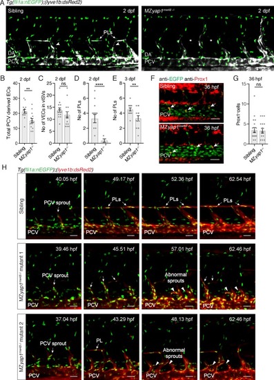
MZ yap1-/- mutants display defects in LEC numbers but not specification.( A) Trunk vasculature (EC nuclei in green, veins and lymphatics in white) of sibling and MZ yap1mw48-/- mutant at 2 dpf. Arrowheads indicate posterior cardinal vein (PCV) sprouts. Asterisks mark absent parachordal LECs (PLs). Dorsal aorta (DA). Scale bars: 50 μm. ( B) Total number of lyve1-positive ECs departing the PCV across 6 somites at 2 dpf. ( C) Number of endothelial cells in venous intersegmental vessels (vISV) across 6 somites at 2 dpf (sibling: 13 ± 0.94, n = 14; MZ yap1-/-: 12 ± 1.33, n = 14; p=0.24 (ns)). ( D) Number of PLs scored across 6 somites at 2 dpf (sibling: 3 ± 0.56, n = 14; MZ yap1-/-: 0.36 ± 0.23, n = 14; p<0.0001 (****)). ( E) Number of PLs scored across 6 somites at 3 dpf (sibling: 4.5 ± 0.20, n = 14; MZ yap1-/-: 3.31 ± 0.36, n = 13; p=0.0075(**)). ( F) Immunofluorescence staining for EC nuclei (green) and Prox1 (red) in sibling and MZ yap1mw48-/- mutants at 36 hpf. Arrows point to Prox1+ LEC progenitors. Scale bars: 30 μm. ( G) Quantification of Prox1 +cells in PCV and CV sprouts scored across 6 somites at 36 hpf (sibling: 3.47 ± 0.65, n = 19; MZ yap1-/-: 3.31 ± 0.61, n = 16; p=0.86 (ns)). ( H) Maximum projection stills from time-lapse Videos from 32 to 65 hpf. Sibling still images show normal lymphangiogenesis with PCV sprouts, PL formation, sprout detachment and PL proliferation (upper panels). MZ yap1mw48-/- mutant one displays abnormal sprouting and looping of PCV sprouts that are retained until the end of the Video (central panels). MZ yap1mw48-/- mutant two also exhibits abnormal sprouting and PCV loop formation but also forms PLs (lower panels). Scale bars: 25 μm. Timelapse imaging began at 32 hpf. |

