FIGURE

Figure 1

ID
ZDB-FIG-190723-1705
Publication
Sarmah et al., 2016 - Zebrafish as a Vertebrate Model System to Evaluate Effects of Environmental Toxicants on Cardiac Development and Function
Other Figures
All Figure Page
Back to All Figure Page
Figure 1

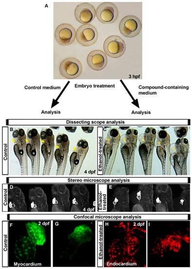
Advantages of use of zebrafish in cardiotoxicity research, which provide enormous information within a short time. (A) Dissecting microscope image of 3 hpf zebrafish embryos showing how easily accessible zebrafish embryos are to treat with chemicals at different developmental stages for desired periods; (B,C) Dissecting microscope images showing normal pericardium in the control embryos (yellow arrow) (B) and pericardial edema phenotype in 4 days post-fertilization (dpf) ethanol-treated zebrafish embryos (yellowstar) to help predict defective cardiogenesis (C); (D,E) Bright field images of Tg(myl7:GFP) embryos showing normal shaped two-chambered heart in control (D) and an almost linear heart in ethanol-exposed embryos (E), confirming heart malformation after ethanol exposure; (F,G) Confocal images of Tg(myl7:nlsKiKGR) embryos showing nuclei of cardiomyocytes in closely apposed bean-shaped atrium and ventricle in control embryos (F), fewer cardiomyocytes are seen in misshapen chambers of ethanol-treated embryos (G); and (H,I) Confocal images of Tg(fli1:EGFP) embryos show endocardial cells in normal endocardium in control embryos (H), fewer endocardial cells are seen in misshapen endocardium of ethanol-treated embryo (I).

Expression Data

Expression Detail
Antibody Labeling
Phenotype Data

Phenotype Detail
Acknowledgments
This image is the copyrighted work of the attributed author or publisher, and ZFIN has permission only to display this image to its users. Additional permissions should be obtained from the applicable author or publisher of the image. Full text @ Int. J. Mol. Sci.