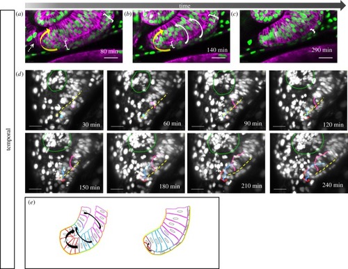
Development of the temporal fissure margin. (a–e) Close up of the developing temporal ventral optic cup domain, (a–c) labelled with tg(bact:H2BGFP) and tg(bact:lyntdTomato), cells from the lens-averted layer of the optic cup (a, bracket) are flowing over the distal rims (a, b white arrows) and in a perpendicular direction (a, b, yellow arrow) over the ventral margin into the lens-facing layer (6 fish in 4 experiments). (c, bracket) Cells in the lens-averted layer (RPE domain) flatten and obtain RPE cell shape. Lateral view, nasal to the left; scale bar, 25 µm. (d) Mosaic nuclear labelling (H2BGFP mRNA injection), maximum projection of 70 optical sections corresponding to 70 µm (z-spacing was 1 µm). Tracking of single cells (from one eye) moving over the distal (magenta and blue, n = 2 for both, respectively) and the ventral distal (red, n = 4) rim (dashed yellow line), respectively, into the lens-facing layer of the prospective neuroretina. Lens marked with green dotted line. Lateral view, nasal to the left; scale bar, 25 µm. (e) Scheme of temporal fissure margin development. Cells move over the distal (magenta), the distal ventral (blue) rims and via a ventral perpendicular flow (red) over the ventral rim (black arrows) from the lens averted into the lens-facing layer.
|
