
Fig. S4
- ID
- ZDB-FIG-190712-2
- Publication
- Yasuda-Yamahara et al., 2018 - FERMT2 links cortical actin structures, plasma membrane tension and focal adhesion function to stabilize podocyte morphology
- Other Figures
- All Figure Page
- Back to All Figure Page
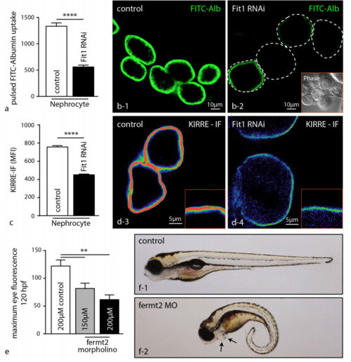
Knockdown of FERMT2 homologs in Drosophilanephrocytes and the zebrafish pronephros. (a–b) RNAi knockdown of the FERMT2 ortholog Fit1(CG14991) was performed in Drosophila nephrocytes (Garland cells) using the RNAi line 46494/GD (Fit1 shows highest similarity to human FERMT2). Pulsed FITC-Albumin uptake assay revealed impaired nephrocyte function in Fit1 knockdown fly (40 cells were analyzed per condition; ****p < .0001). (c–d) The amount of the slit diaphragm marker Kirre was analyzed by anti-Kirre 126i immunofluorescence (IF) staining. MFI (mean fluorescence intensity) quantification revealed a significant reduction (40 cells were analyzed per condition; ****p < .0001). (e–f) Analysis of fermt2-knockdown in zebrafish larvae revealed edema (pericardial effusion, arrows) and loss of high molecular weight proteins from the circulation indicating impaired pronephros function in fermt2 morpholinos injected zebrafish embryos (Maximum eye fluorescence of 30 control, 25,150 μM fermt2 and 12,200 μM fermt2 morpholinos was measured at 120hpf; **p < .01). |
| Fish: | |
|---|---|
| Knockdown Reagent: | |
| Observed In: | |
| Stage: | Day 5 |