
Fig. 3
- ID
- ZDB-FIG-190705-6
- Publication
- Narboux-Neme et al., 2019 - Posterior axis formation requires Dlx5/Dlx6 expression at the neural plate border
- Other Figures
- All Figure Page
- Back to All Figure Page
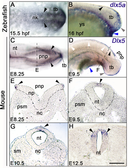
Dlx5 expression analysis during zebrafish and mouse posterior neurulation. (A-D) Whole-mount in situ hybridization for dlx5a and Dlx5; (A, C) dorsal and (B, D) lateral views of the posterior axis of 15.5 hpf and 16 hpf zebrafish (A-B) and E8.25 and E9.5 mouse embryos (C-D). (E-H) In situhybridization for Dlx5 on coronal cryosections at the levels indicated by the dashed lines in (C, D and S1 Fig). In zebrafish embryos, dlx5a transcripts are detected in NPB cells along neural keel at 15.5 hpf and at the dorsal midline at 16 hpf (A-B, black arrowheads). During mouse posterior neurulation, Dlx5 is expressed in NPB cells surrounding the posterior neuropore and along the dorsal midline of the neural tube after neural tube closure (C-H, black arrowheads). In both species, Dlx5 is also detected in the ventral ectodermal ridge of the tail bud and at the cloacal level (grey and blue arrowheads respectively in B, D) (n>10 for each conditions). Abbreviations: nc, notochord; nk, neural keel; np, neural plate; nt, neural tube; pnp, posterior neuropore; psm, presomitic mesoderm; sm, somitic mesoderm; tb, tail bud; ys, yolk sac. Scale bar in H for A, H 50 μm, for B, E-G 75 μm, for C 150 μm, for D 200 μm. |
| Gene: | |
|---|---|
| Fish: | |
| Anatomical Terms: | |
| Stage Range: | 10-13 somites to 14-19 somites |