
Fig. 5
- ID
- ZDB-FIG-190701-24
- Publication
- Singh et al., 2019 - High glucose levels affect retinal patterning during zebrafish embryogenesis
- Other Figures
- All Figure Page
- Back to All Figure Page
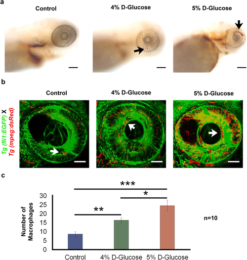
Increased haemoglobin presence and retinal blood vessel leakage following pulsatile high-glucose exposure from 3 hpf until 5 dpf. (a) The increased presence of haemoglobin in the retina (indicated with black arrows) was detected in WT embryos exposed to 4 and 5% D-Glucose in a pulsatile manner from 3 hpf until 5 dpf following O-dianisidine staining indicating increased accumulation of hemoglobin in the retinal blood vessels, which is indicative of leaky retinal blood vessels. Control WT embryos were maintained in the vehicle (E3) only from 3 hpf until 5 dpf under standard conditions. (n = 5). Scale bar, 50 µm. (b) Retinal blood vessel leakage following pulsatile D-Glucose exposure. Tg (fli1:EGFP) X Tg (mpeg:DsRed) embryos were exposed to a fluctuating 4 and 5% D-Glucose treatment from 3 hpf until 5 dpf. Scale bar, 50 µm. (c) Statistical analysis of Tg (fli1:EGFP) X Tg (mpeg:DsRed) exhibited an increase in the number of macrophages (indicated by white arrows) in the retina at 5 dpf, indicating blood vessel leakage. Error bars indicate mean ± s.e.m.; Statistical differences were computed using two-tailed student’s t-test and are indicated as *p = 0.03, **p = 0.007, ***p < 0.001; (n = 10).
|
| Gene: | |
|---|---|
| Fish: | |
| Condition: | |
| Anatomical Term: | |
| Stage: | Day 5 |
| Fish: | |
|---|---|
| Condition: | |
| Observed In: | |
| Stage: | Day 5 |