
FIGURE
Fig. 2
Fig. 2
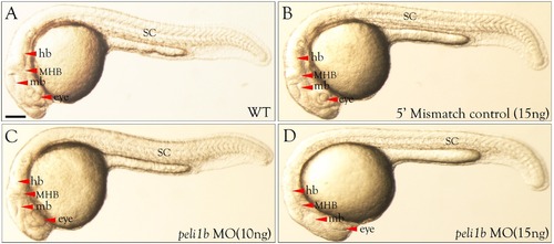
Morphological defects in zebrafish embryos after knockdown of peli1b. All images were taken at 24 hpf. (A) WT, (B) 5′ mismatch control, (C) peli1b MO (10 ng), and (D) peli1b MO (15 ng). Morpholino and 5′ mismatch control were injected at the 1-cell stage in synchronized embryos. (A–D) Lateral view of the embryos showing the phenotype after 10 ng or 15 ng injection of peli1b antisense nucleotidemorpholino. Abbreviations: NT, neural tube; SC, spinal cord; fb, forebrain; mb, midbrain; hb, hindbrain; MHB, midbrain–hindbrain boundary. (n = 3). Scale bar- 50 μm. |
Expression Data
Expression Detail
Antibody Labeling
Phenotype Data
| Fish: | |
|---|---|
| Knockdown Reagent: | |
| Observed In: | |
| Stage: | Prim-5 |
Phenotype Detail
Acknowledgments
This image is the copyrighted work of the attributed author or publisher, and
ZFIN has permission only to display this image to its users.
Additional permissions should be obtained from the applicable author or publisher of the image.
Reprinted from Gene, 694, Kumar, A., Anuppalle, M., Maddirevula, S., Huh, T.L., Choe, J., Rhee, M., Peli1b governs the brain patterning via ERK signaling pathways in zebrafish embryos, 1-6, Copyright (2019) with permission from Elsevier. Full text @ Gene