
Fig. 3
- ID
- ZDB-FIG-190628-31
- Publication
- Guesmi et al., 2018 - Dual-color deep-tissue three-photon microscopy with a multiband infrared laser
- Other Figures
- All Figure Page
- Back to All Figure Page
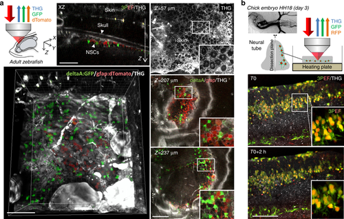
a In vivo imaging through the skull in adult zebrafish telencephalon. The figures show representative XY, XZ, and 3D views for a volume encompassing two labeled cell populations: red dTomato-labeled radial glia and green GFP-labeled neural stem cells expressing the deltaA neurogenic gene (see Materials and methods section). Simultaneously acquired THG signals provide additional label-free contextual information (skin and skull morphology, blood vessels, lipid accumulations). See also Movies 7 and 8 and related information. b Simultaneous dual-color 3PEF and THG imaging of developing chick embryo spinal cord tissue (stage E3) expressing cytoplasmic GFP labeling and nuclear RFP labeling. The images are extracted from a 2-h-long experiment and illustrate developmental processes such as cell migration and process formation. See also Movies 5 and 6 and related information. Scale bars, 100 µm |