
FIGURE
Fig. S1
- ID
- ZDB-FIG-190626-34
- Publication
- Camargo-Sosa et al., 2019 - Endothelin receptor Aa regulates proliferation and differentiation of Erb-dependent pigment progenitors in zebrafish
- Other Figures
- All Figure Page
- Back to All Figure Page
Fig. S1
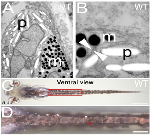
Melanocytes and iridophores in the WT yolk sac stripe are consistently separated from each other by double membranes.Transmission electron photomicrographs of melanocytes and iridophores in the WT yolk sac stripe ectopic pigment cells in pde mutants. A and B show two examples of melanosomes (m) and reflecting. platelets (p) separated by a double membrane (white arrowheads). Bright-field image of ventral view (C) and close up of the area in the red box (D) of WT fish, shows yolk sac stripe. Continuous layer of iridophores is indicated by white arrowhead in D, closely associated black melanocytes forming contiguous layer immediately dorsal to iridophores is indicatted by red arrowhead. Scale bars = 500 μm (C) and 100 μm (D). |
Expression Data
Expression Detail
Antibody Labeling
Phenotype Data
Phenotype Detail
Acknowledgments
This image is the copyrighted work of the attributed author or publisher, and
ZFIN has permission only to display this image to its users.
Additional permissions should be obtained from the applicable author or publisher of the image.
Full text @ PLoS Genet.