
Fig. S3
- ID
- ZDB-FIG-190626-19
- Publication
- Goudarzi et al., 2019 - Fluid dynamics during bleb formation in migrating cells in vivo
- Other Figures
- All Figure Page
- Back to All Figure Page
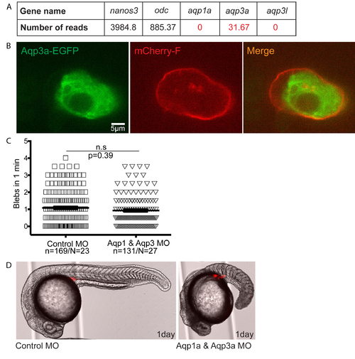
(A) The table shows the number of reads of specific mRNAs in PGCs at 7 hours post fertilization (hpf), based on the microarray sequencing data from [25]. The data includes a PGC specific gene (nanos3), a housekeeping gene (odc) and aquaporin 1 and 3 isoforms. (B) Subcellular localization of Aqp3-GFP expressed in the PGCs employing the 3’-untranslated region of nanos3. (C) A graph showing the blebbing activity of PGCs in embryos injected with either 800μM of control morpholino or with 400μM Aqp1a morpholino + 400μM Aqp3a morpholino. N is the number of embryos and n represents the number of cells analyzed. The graph shows the mean and the standard deviation. (D) A low magnification image showing the morphology of 1-day old embryos treated with control and aqp 1 & 3 morpholinos. The PGCs are labeled in red. Scale bar = 5 μm. |