
Fig. 2
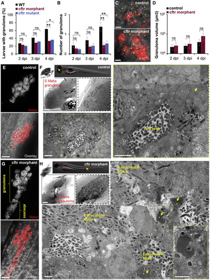
Absence of CFTR Impairs M. abscessus Infection-Mediated Granuloma Integrity and Maintenance (A–D) WT, cftr mutants, and cftr morphants were i.v. infected with the S variant of Mabs expressing tdTomato and monitored using confocal microscopy for granuloma formation, number, and size (n = 35–45; data are plotted as mean ± SEM from three experiments). (A and B) Kinetics of granuloma formation in whole embryos over 4 days of infection (A) and associated number of granuloma per animal (B). (C) Confocal images showing a representative early Mabs granuloma in 3 dpi larvae. Scale bars, 10 μm. For a similar number of infected phagocytes(asterisk) in a granuloma, the CF granuloma contains a higher bacterial burden than the WT granuloma. (D) Granuloma volume analysis in whole larvae over 4 days of infection. (E–H) Control larvae or cftr morphants infected with Mabs S expressing tdTomato observed by confocal microscopy and EM for granuloma at 4 dpi. (E and G) Representative confocal imaging of a granuloma within the spinal cord of an infected animal showing the development of a compact and organized phagocyte aggregate moderately infected in control fish (E) compared with a cftr morphant harboring a heavily loaded granuloma whose disruption leads to bacterial spread characterized by abscess formation (G). Scale bars, 10 μm. (F) EM showing a sagittal section through a WT Mabs granuloma. Overview image of granuloma (arrow) into the brain of infected WT (top; scale bar, 50 μm). Representative EM image of a compact and well-organized WT granuloma (bottom and right; processed as stitching of micrographs; scale bars, 2 μm) showing a stable mycobacterial-containing structure with a central necrotic area and a typical dense region of cellular debris and surrounded by numerous infected phagocytes (asterisk) and giant cells (arrow). (H) EM showing a sagittal section through a Mabs granuloma-like lesion in absence of CFTR. Overview image of granuloma in the spinal cord (arrow) of infected cftr morphant (top; scale bar, 25 μm). Representative EM image of a CFTR-depleted granuloma (bottom and right; processed as stitching of micrographs; scale bar, 2 μm) showing a necrotic structure with replicating bacteria and numerous dead infected phagocytes harboring a typical apoptotic nucleus (asterisk) with chromatin“superaggregation.” Most bacilli stay extracellular. |