
Fig. 6
- ID
- ZDB-FIG-190620-49
- Publication
- Huo et al., 2019 - Transcriptomic profiles of tumor-associated neutrophils reveal prominent roles in enhancing angiogenesis in liver tumorigenesis in zebrafish
- Other Figures
- All Figure Page
- Back to All Figure Page
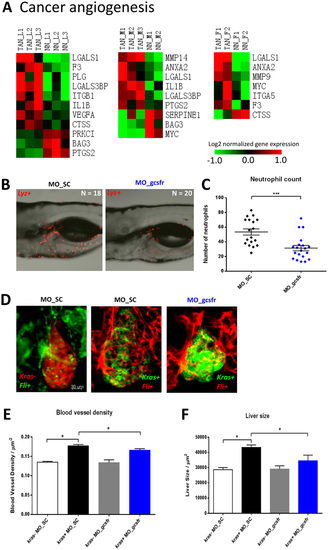
Expression of cancer angiogenesis genes in TANs and effects of neutrophil depletion on liver tumorigenesis and angiogenesis. (A) Heatmap representation of expression of cancer angiogenesis genes extracted from DEGs of TANs. The color codes represent fold changes of expression of each gene in each sample relative to the mean expression of all samples. The list of cancer angiogenesis genes was obtained based on IPA knowledge datbase. (B) Representative confocal images of 6-dpf lyz+ larvae injected with different morpholinos: MO_SC (left, n = 18) or MO_gcsfr (right, n = 20). (C) Numbers of neutrophils in the middle body after injection of morpholinos. Neutrophils were counted in the the middle body surrounding the liver region (from the posterior edge of eye to posterior edge of swimbladder). (D) Representative 3D confocal images of 6-dpf kras−/fli+ and kras+/fli+ larvae injected with MO_SC or MO_gcsfr. For fli+ larvae, both Tg(fli1a::GFP) (left) and Tg(fli1a::RFP) (middle and right) were used. Kras- larvae were LiPan strain (left) with DsRed expression in the liver. (C and D) Quantification of blood vessel density in the liver area (E) and liver size (F) after morpholino injection. N >20 in each group. Statistical significance: *p < 0.05.
|
| Gene: | |
|---|---|
| Fish: | |
| Knockdown Reagent: | |
| Anatomical Term: | |
| Stage: | Day 6 |
| Fish: | |
|---|---|
| Condition: | |
| Knockdown Reagent: | |
| Observed In: | |
| Stage: | Day 6 |