
Fig. 5
- ID
- ZDB-FIG-190613-17
- Publication
- Ferguson et al., 2019 - The catalytic activity and secretion of zebrafish RNases are essential for their in vivo function in motor neurons and vasculature
- Other Figures
- All Figure Page
- Back to All Figure Page
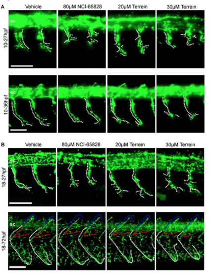
Axonal defects in zebrafish embryos treated with NCI-65828 and terrein. Immunostaining for Znp1 in Fli:GFP zebrafish treated with NCI-65828 or terrein from (A) 10 hpf or (B) 18 hpf. Znp1 staining shows defects in the extension and branching of the caudal primary motor neuron (CaP, white dotted lines) extending from the spinal cord (sc). In addition to shortened axons, additional aberrant branching can be seen in treated embryos at positions more proximal to the spinal cord when compared to untreated, which branch only at the distal regions of the axon. Axons in embryos treated at 10 hpf also appear to loop and branch anteriorly at 36 hpf frequently while untreated continue to extend towards the posterior. Treatment from 18 hpf still results in reduced axon length at 27 hpf but to a lesser degree than those treated at 10 hpf. Aberrant branching is still present but with increased branching at the distal tip. By 72 hpf CaP axons appear positioned normally in treated and untreated alike. Observations from 10 embryos in two replicates show significant effects. Detailed quantification can be found in Figure S1. Defects are still seen in the rostral and medial primary motor neurons (RoP and MiP, red and blue dotted lines) and secondary motor neurons (not shown). See Figure S2 for quantification. Scale bars 50 µm.
|