
Fig. 2
- ID
- ZDB-FIG-190604-43
- Publication
- Zhao et al., 2019 - Endocardial Notch Signaling Promotes Cardiomyocyte Proliferation in the Regenerating Zebrafish Heart through Wnt Pathway Antagonism
- Other Figures
- All Figure Page
- Back to All Figure Page
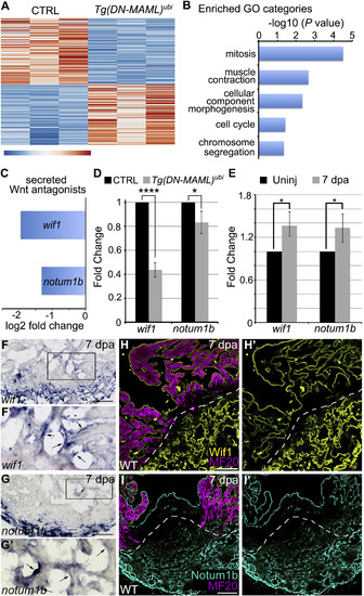
Identification of Secreted Wnt Signaling Antagonists as Candidate Notch Targets in the Endocardium during ZebrafishHeart Regeneration (A) Heatmap showing 254 differentially expressed genes (p < 0.0001) in wild-type (CTRL; n = 3 replicates) and Tg(hsp70l:DN-MAML) (n = 3 replicates) hearts on 5 dpa with daily heat shocking. (B) Bar graph showing −log10 p values for GO terms significantly overrepresented in the downregulated gene category. (C) Bar graph showing the log2 fold-change values for transcripts encoding wif1 and notum1b from (A). (D) Bar graph showing the relative expression levels of wif1 and notum1b in hearts of CTRL and hsp70l:DN-MAML animals on 5 dpa with daily heat shocking as measured by qPCR. (E) Bar graph showing the relative expression levels of wif1 and notum1b in hearts of wild-type animals without injury and on 7 dpa as measured by qPCR. (D and E) 3 technical replicates of each of 3 biological replicates were performed. Statistical significance was determined using a Student’s t test. Error bars: ±1 SD. ∗∗∗∗p < 0.0001. ∗p < 0.05. (F–G′) In situ hybridization for wif1 and notum1b in cardiac sections of wild-type hearts on 7 dpa. Boxed regions in (F) and (G) are enlarged in (F′) and (G′). Arrowheads highlight endocardial signals. 6–8 sections from 4–6 hearts were analyzed. Little to no variation was observed. (H–I′) Immunohistochemical analysis of Wif1 (H and H′; yellow) and Notum (I and I′; cyan) in cardiac sections of wild-type hearts on 7 dpa costained with the MF20 antibody (magenta) to visualize myocardium. Single confocal slices are shown. Dashed lines highlight approximate amputation planes. 6–8 sections from 4–6 hearts were analyzed. Little to no variation was observed. Scale bars: 50 μm. |