
Fig. 1
- ID
- ZDB-FIG-190528-6
- Publication
- Blokhina et al., 2019 - The telomere bouquet is a hub where meiotic double-strand breaks, synapsis, and stable homolog juxtaposition are coordinated in the zebrafish, Danio rerio
- Other Figures
- All Figure Page
- Back to All Figure Page
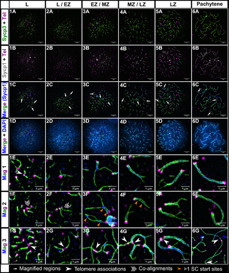
Homologous chromosome synapsis in male zebrafish meiosis I. 1A-6A: Loading of the chromosome axis protein, Sycp3 (green), initiates near the telomeres (Tel; magenta) in the bouquet and extends inward from both ends of the chromosome during meiotic prophase progression at the indicated stages (leptotene (L); leptotene to zygotene transition (L/EZ); early to mid-zygotene (EZ/MZ); mid to late zygotene (MZ/LZ); late zygotene (LZ); pachytene). 1B-6B: Sycp3 loading is closely followed by the initiation of SC formation, marked by Sycp1 (gray in row B to highlight features of the SC, blue in following rows)). 1C-6C: Merge of Sycp3, Sypc1 and Tel staining. 1D-6D: Merge of all signals plus DNA staining with DAPI. 1E-6G: Magnifications of three different regions (Mag 1, Mag 2, and Mag3) for each of the corresponding panels above; each magnified region is specified in images 1C-6C by long arrows. 1E, 1G-6G: Telomere associations are seen at all stages (arrowheads). 2G: Telomere association between three telomeres. 1F-2F: Short stretches of pre-synaptic co-alignment (triple arrows). 3F-4F: Chromosomes with more than one synapsis initiation site (orange arrows). 1A-6D scale bar = 5 μm. 1E-6G scale bar = 1 μm.
|
| Antibodies: | |
|---|---|
| Fish: | |
| Anatomical Terms: | |
| Stage: | Adult |