|
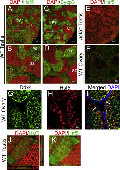
Hsf5 Protein Showed Similar Localization Pattern in Spermatocytes to a Known Spermatocyte Marker Sycp3 (A and B) Representative images show immunostaining with anti-Hsf5 antibodies on adult WT testis sections (B is a zoomed-in image of A). (C and D) Adjacent sections were immunostained with anti-Sycp3 antibodies (D is a zoomed-in image of C), and nuclei were counterstained with DAPI. Strong Hsf5 and Sycp3 signals were detected in spermatocytes, while weak Hsf5 signals were seen in spermatogonia, spermatids and spermatozoa. (E) Immunostaining with anti-Hsf5 antibody did not show significant signals in hsf5−/− testis. (F) Immunostaining suggests low levels of Hsf5 in WT ovary. (G–I) Representative images show immunostaining of adult WT ovaries from tg(ddx4:ddx4-egfp) transgenic zebrafish with anti-Ddx4 (green; G) and anti-Hsf5 (red; H) antibodies, respectively, and merged images showing signals from both antibodies (I). Hsf5 localization pattern follows that of Ddx4 and does not stain the gonadal somatic cells. White arrows indicate the outer layer of oocytes. (J) Localization of Hsf5 protein (green) as granule-like structures in the nucleus of primary spermatocytes; orthogonal views are presented to emphasize the presence of foci in the nucleus (red). (K) Maximum-intensity Z-projection of images. For (A)–(K), n = 3 biological replicates in each group. Scale bars: 50 μm in (A), (C), (G), (H), and (I); 30 μm in (B) and (D); 400 μm in (E); 100 μm in (F); and 5 μm in (G), (H), and (K). See also Figure S4.
|