|
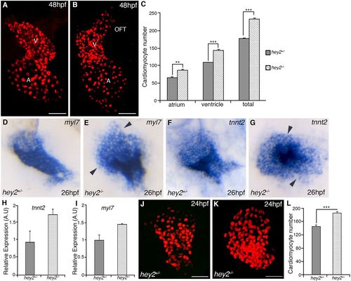
Loss of Hey2 results in heightened cardiomyocyte number. (A,B) Confocal images showing cardiomyocyte nuclei at 48 hpf in hey2 heterozygous (A) and mutant (B) embryos. (C) Counts of cardiomyocyte numbers in the atrium and ventricle of embryos at 48 hpf (N=3, n=6 per condition). (D-G) RNA in situ hybridization analysis for expression of myl7 (D,E) and tnnt2 (F,G) at 26 hpf. Arrowheads indicate ectopic cells outside the cardiac cone in hey2 mutants. (H,I) Quantitative RT-PCR analysis of tnnt2 (H) and myl7 (I) transcript levels at 26 hpf (gene expression normalized to β-actin, fold difference relative to control; N=3, n=3). (J,K) Confocal images of cardiomyocyte nuclei in 24 hpf hey2 heterozygous (J) and hey2 mutant (K) Tg(myl7:nlsDsRedExpress) embryos. (L) Total cardiomyocyte number at 24 hpf in hey2 heterozygous and mutant embryos (N=3, n=5 per condition). Scale bars: 50 µm. Data are mean±s.e.m.; **P<0.01; ***P<0.001. A, atrium; V, ventricle; OFT, outflow tract.
|