
Fig. 3
- ID
- ZDB-FIG-190118-18
- Publication
- Love et al., 2018 - Vangl2-dependent regulation of membrane protrusions and directed migration requires a fibronectin extracellular matrix
- Other Figures
- All Figure Page
- Back to All Figure Page
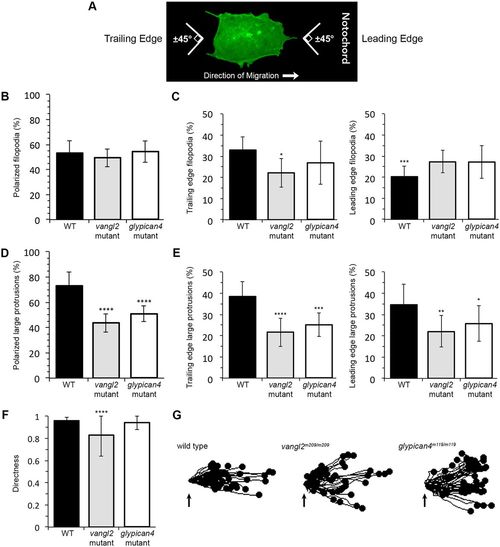
Membrane protrusion polarity and directed migration in vangl2 and glypican4 mutant embryos. (A) Wild-type (WT) ectodermal cell image illustrating polarized protrusion angles. (B,C) Quantitation of the total percentages of polarized filopodia (B) and filopodia localized at the leading versus trailing edge (C) in WT (n=11 cells, 8 embryos), vangl2m209/m209 (n=10 cells, 7 embryos) and glypican4m119/m119 (n=11 cells, 5 embryos). (D,E) Quantitation of polarized large protrusions (D) and large protrusions localized at the leading versus trailing edge (E) in WT (n=12 cells, 8 embryos), vangl2m209/m209 (n=10 cells, 7 embryos) and glypican4m119/m119 (n=12 cells, 5 embryos). (F) Directed migration values in WT (n=50 cells, 11 embryos), vangl2m209/m209 (n=51 cells, 13 embryos) and glypican4m119/m119 (n=50 cells, 8 embryos). (G) Schematic of the migration paths of individual ectodermal cells. Origins (arrows) standardized for comparison. Dorsal is to the right. Data are mean±s.d. *P<0.05, **P<0.01, ***P<0.001, ****P<0.0001; P values are versus WT, except WT leading edge P value which is versus WT trailing edge; one-way ANOVA significance test followed by Tukey HSD post-hoc tests. |