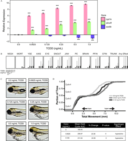
Analysis of the concentration–response effects of developmental exposure (1-h exposure at 6hpf ) to TCDD on gene expression at 48hpf and morphological malformations at 120hpf . (A) qRT-PCR relative expression of cyp1a, slincR, and sox9b transcripts in 48-hpf wild-type embryos exposed to 0, 0.625, 0.125, 0.25, 0.5, or 1.0ng/mL TCDD. For all assays, 0.1% DMSO served as the vehicle control and is listed as 0ng/mL TCDD. Expression values were analyzed with the 2−ΔΔCT method and normalized to β-actin using the 0ng/mL TCDD concentration as the calibrator. Each experimental unit represents a pool of 20 embryos, and each treatment group included four biological replicates ( n=4 ). The data were log2-transformed and analyzed using a one-way ANOVA with a Dunnett post hoc test ( p<0.001 in comparison with 0ng/mLTCDD=*** ). All error bars indicate standard error of the mean. (B) Evaluation of 17 physical malformations at 120-hpf on wild-type zebrafish exposed to six concentrations of TCDD ( 0–1ng/mL ) across two 96-well plates. Nonsignificant malformations (otic vesicle, somite, circulation, pigmentation, swim bladder, notochord distortion, and alterations in touch response) are excluded. The horizontal axis displays the TCDD concentrations tested, and the malformation examined is listed above each box. The incidence across all replicates is plotted as stacked points. For each malformation, the stacked points exceeding the binomial significance threshold are represented in light gray (top stack). The data were analyzed using a Fisher’s exact test with a Bonferroni correction for multiple comparisons ( n=32 , p<0.01 ). (C) Representative lateral images of 120-hpf zebrafish for each concentration of TCDD tested. The bar in top left corner indicates 100μM . (D) Larval photomotor response (LPR) in 120-hpf wild-type embryos developmentally exposed to 0, 0.0625, or 0.125ng/mL TCDD equally divided across two 96-well plates using the ViewPoint ZebraBox larvae screening system. For each concentration of TCDD, the overall area under the curve was analyzed for the last 3 light:dark cycles in comparison with 0ng/mL TCDD using a Kolmogorov-Smirnov test ( n=32 , p≤0.01 ).
|
