
Fig. 3
- ID
- ZDB-FIG-181127-44
- Publication
- Genoud et al., 2018 - Fast Homogeneous En Bloc Staining of Large Tissue Samples for Volume Electron Microscopy.
- Other Figures
- All Figure Page
- Back to All Figure Page
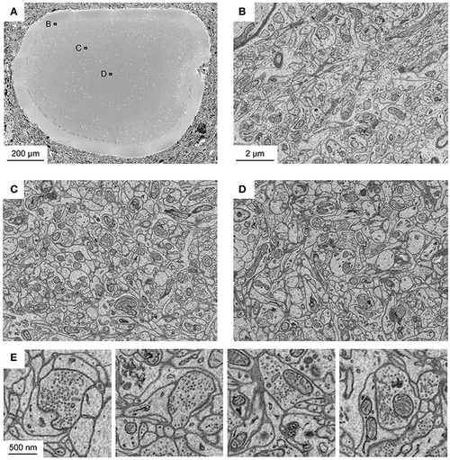
Application of fBROPA to the adult zebrafish brain. (A) Section through the tectum near the location where the diameter is maximal (1.1 mm). Image is a mosaic of 6 × 6 tiles. (B–D) Three images acquired at different depths. Approximate locations of images are indicated by outlines in (A). (E) Examples of images showing synapses (5 nm pixel size). Vesicle pools close to the presynaptic membrane and a thickening of both membranes are visible. Synapse detection can be performed in 3D as shown in Supplementary Data S1 (movie). Note uniformly high contrast. The partial damage on the right side of the tectum occurred during dissection and is unrelated to fixation or staining. |