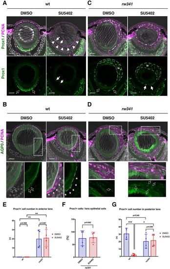
Ectopic lens fiber differentiation in rw341 mutants is independent of FGF signaling. (A) Prox1 and PCNA expression in wild-type lenses. DMSO treatment does not affect PCNA and Prox1 expression, which are normally expressed in lens epithelium and newly differentiating lens fiber cells (open arrows), respectively. SU5402 treatment inhibits Prox1 expression. Almost all cells along the posterior margin of the lens fiber region express only PCNA (arrowheads). A few weakly Prox1-expressing cells are observed inside PCNA-positive posterior marginal cells (filled arrows). (B) AQP0 and PCNA expression in wild-type lenses. Bottom panels indicate higher magnification of outlined areas in the upper panels. DMSO treatment does not affect AQP0 expression. A few peripheral PCNA-positive cells express AQP0 (open arrows). In SU5402 treatment, almost all PCNA-positive posterior marginal cells did not express AQP0 (asterisks). (C) Prox1 and PCNA expression in rw341 mutant lenses. DMSO treatment does not change Prox1 expression in the posterior lens fiber region (open arrows) as well as the anterior multilayered epithelium (le). SU5402 treatment also did not inhibit Prox1 expression in either the anterior lens epithelium (le) or the posterior lens fiber region (filled arrows). (D) AQP0 and PCNA expression in rw341 mutant lenses. Bottom panels indicate higher magnification of outlined areas in the upper panels. AQP0-positive cells are detected in the anterior multilayered lens epithelium of both DMSO- and SU5402-treated rw341 mutants (open arrows). (E) Number of Prox1-positive cells in the anterior lens epithelium per lens section. There are no Prox1-positive cells in wild-type lens epithelium treated with either DMSO or SU5402. SU5402 treatment did not reduce Prox1-positive cells in rw341 mutants. (F) Percentage of Prox1-positive cells relative to total lens epithelial cells. SU5402 treatment did not reduce the fraction of Prox1-positive cells in rw341 mutants. (G) Number of Prox1-positive cells in the posterior lens area per lens section. SU5402 treatment drastically reduces Prox1-positive cells in wild type, but did not in rw341 mutants. (E-G) Data are mean±s.d. (E,G) Two-way ANOVA. (F) Student's t-test: *P<0.05, **P<0.01, ***P<0.005. Scale bars: 20 µm (10 µm in higher magnification images in B,D).
|
