
Fig. 7
- ID
- ZDB-FIG-181120-21
- Publication
- Fernando et al., 2018 - Diphlorethohydroxycarmalol Isolated from Ishige okamurae Represses High Glucose-Induced Angiogenesis In Vitro and In Vivo.
- Other Figures
- All Figure Page
- Back to All Figure Page
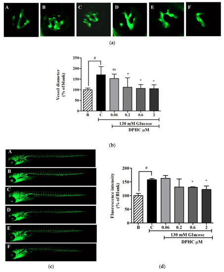
DPHC inhibited high glucose-induced dilation in hyaloid retinal vessel diameter and the whole body vessel formation in the zebrafish transgenic (flk:EGFP) embryo. A: 0 mM glucose + 0 μM DPHC; B: 130 mM glucose + 0 μM DPHC; C: 130 mM glucose + 0.06 μM DPHC; D: 130 mM glucose + 0.2 μM DPHC; E: 130 mM glucose + 0.6 μM DPHC; F: 130 mM glucose + 2 μM DPHC. (a) Images of hyaloid retinal vessels of the zebrafish transgenic (flk:EGFP) embryo taken with florescence microscopy (10×). (b) Quantification of the hyaloid retinal vessel diameter. (c) Images of the zebrafish transgenic (flk:EGFP) embryo’s whole body taken with florescence microscopy (4×). (d) Quantification of the whole body florescence intensity. The effect of 130 mM of glucose is compared with the blank (130 mM glucose + 0 μM DPHC), # p ˂ 0.05. The effect of DPHC against high glucose-induced vessel growth is normalized to the control (130 mM glucose + 0 μM DPHC); ns, not significant, * p ˂ 0.05. |
| Fish: | |
|---|---|
| Conditions: | |
| Observed In: | |
| Stage: | Protruding-mouth |