
Fig. 6
- ID
- ZDB-FIG-181115-11
- Publication
- Formella et al., 2018 - Real-time visualization of oxidative stress-mediated neurodegeneration of individual spinal motor neurons in vivo
- Other Figures
- All Figure Page
- Back to All Figure Page
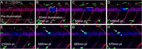
Microglia migrate towards the site of KR activation. (A-H) Time lapse imaging of a zebrafish expressing green fluorescent microglia (mpeg1:EGFP), and MNs labelled in blue (mnx1:mTagBFP) and expressing red KR (mnx1:MLS-KillerRed) throughout the spinal cord. Post-illumination (pI, B, white dotted line) KR fluorescence was significantly reduced while TagBFP fluorescence remained unaffected. Green fluorescent microglia within close proximity extended its processes towards to the KR activation site within the first two hours (C-D), seemingly inspecting light-targeted MN-bodies. Microglia subsequently moved away from the illumination site (E). Approximately 9 h post-illumination, microglia were again observed at the illumination site (F). These microglia underwent characteristic morphological changes (amoeboid body) and remained at the KR activation site for several hours (G-H). Notably, near the site of microglia activity a TagBFP+ve MN disappeared, conceivably indicating its death due to KR activation (Supplementary Video 4). Scale bar 50 µm. |