
Fig. 2
- ID
- ZDB-FIG-181101-2
- Publication
- Willoughby et al., 2012 - Generation of a genetically encoded marker of rod photoreceptor outer segment growth and renewal
- Other Figures
- All Figure Page
- Back to All Figure Page
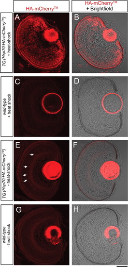
Expression of HA-mCherryTM in the eye of a 69.5 hpf larva at 1.5 hours hpHS (heat shocked at 68 hpf). (A, C, E, G) Single confocal z-section images of an eye labeled with anti-HA antibodies. (B, D, F, H) Single confocal z-section image of an eye labeled with anti-HA antibodies merged with DIC-like image. (A, B) Heat-shocked Tg(hsp70:HA-mCherryTM) larva shows ubiquitous expression of HA-mCherryTM in retinal cells. (C, D) Heat-shocked wild-type eye shows only weak autofluorescence. (E, F) In the absence of heat-shock, an eye of a Tg(hsp70:HA-mCherryTM) larva shows only weak expression of HA-mCherryTM in a small number of amacrine cells (arrows). (G, H) An eye of a wild-type larva shows weak autofluorescence in the absence of heat-shock. Scale bar: A–H, 50 µm. |