
Fig. 3
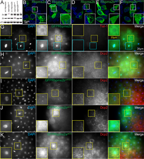
rbpms2 mutant allele stability and localization activity in somatic cells. (A) RT-PCR to detect rbpms2a and rbpms2b maternal transcripts. Pink asterisks indicate likely nonsense mediated decay of mutant allele transcripts. (B-F) Wild-type GFP-Rbpms2a/b (B, D) and GFP-Rbpms2bsa9329 (F) localize to granules in HEK 293 cells, while GFP-Rbpms2aae30 and GFP-Rbpms2bae32 are not localized. (G-K) Zebrafish blastula cells expressing GFP-Rbpms2 fusions. (G,H) Wild-type GFP-Rbpms2b localizes near the nucleus (H), is apparently associated with the centrosome/spindle in some cells, and (G) is in granules that are not positive for the stress granule marker Tial-1. (H) A subset of GFP-Rbpms2b positive granules are positive for the p-body marker Dcp2 (open arrowheads). (I) GFP-Rbpms2bsa9329 localization to the centrosome/spindle but not granules. (J) GFP-Rbpms2bae32 and (K) GFP-Rbpms2aae30 localization. Insets show magnified views of the highlighted cells. Images are representative slices from Z-stacks of sphere stage embryos viewed from the animal pole. |
| Genes: | |
|---|---|
| Fish: | |
| Anatomical Term: | |
| Stage: | 4-cell |
| Fish: | |
|---|---|
| Observed In: | |
| Stage: | 4-cell |