
Fig. 5
- ID
- ZDB-FIG-181019-16
- Publication
- Campbell et al., 2018 - A high content, small molecule screen identifies candidate molecular pathways that regulate rod photoreceptor outer segment renewal
- Other Figures
- All Figure Page
- Back to All Figure Page
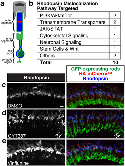
Rhodopsin mislocalizes following treatment with JAK1/2 and microtubule inhibitors. (a) Rhodopsin normally localizes to the ROS and is mislocalized when detected in the inner segment, cell body, or synaptic region with Rhodopsin immunolabeling. (b) Pathways targeted by compounds that lead to Rhodopsin mislocalization. (c) Rhodopsin localizes to the ROS in DMSO treated larva. (d) Rhodopsin is mislocalized to the inner segment, as well as some puncta in the cell body (arrows), following treatment with the JAK1/2 inhibitor CYT387. (e) Rhodopsin is mislocalized to the inner segment and cell body following treatment with Vinflunine, which disrupts microtubules. Merged images show GFP-expressing rods in green, HA-mCherryTM stripe in red, and Rhodopsin immunolabeling in blue. Scale bar is 10 μm. |