
Fig. 1
- ID
- ZDB-FIG-181003-2
- Publication
- Loynes et al., 2018 - PGE2 production at sites of tissue injury promotes an anti-inflammatory neutrophil phenotype and determines the outcome of inflammation resolution in vivo.
- Other Figures
- All Figure Page
- Back to All Figure Page
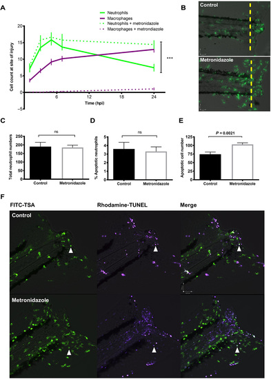
Macrophage clearance of apoptotic cells is necessary for successful resolution of neutrophilic inflammation in an in vivo zebrafish model. (A) Neutrophil and macrophage counts at the wound in triple transgenic Tg(cfms:Gal4)i186;Tg(UAS:nfsB-mCherry)i149;Tg(mpx:EGFP)i114 larvae in the presence or absence of metronidazole. Neutrophil numbers are significantly higher at the wound in the absence of macrophages at 24 hpi, ***P < 0.0001. Statistics: Two-tailed nonpaired t test comparing neutrophil counts at 24 hpi with or without macrophage present. All data are n = 18 from three individual experiments plotted as means ± SEM. (B) Representative photomicrographs at 24 hpi showing increased neutrophil numbers at the site of injury in metronidazole-treated larvae compared to control. Wound area classed as area to the right of the yellow dashed line. Images were taken using ×10 magnification on a TE2000U inverted microscope (Nikon). (C) Total neutrophil numbers are not affected in the absence of macrophages. Data are n = 17 individual larvae. (D) Dual fluorescein isothiocyanate (FITC)–TSA and Rhodamine-TUNEL–positive cell counts in 8 dpf larvae at 24 hpi in the presence or absence of metronidazole show no significant difference (ns) in the percentage of apoptotic neutrophils. (E) Total apoptotic cell counts of TUNEL-positive cells in 8 dpf larvae at 24hpi. Macrophage-depleted larvae have significantly more apoptotic bodies at the wound, P = 0.0021. All data are presented as means ± SEM, n = 12, from two individual experiments for (D) and (E). (F) Representative photomicrographs of 8 dpf larvae at 24 hpi treated with either dimethyl sulfoxide (DMSO) control or metronidazole and dual-stained with FITC-TSA to label neutrophils and Rhodamine-TUNEL to label apoptotic cells at the wound (white arrow head). Images were taken using ×10 magnification on a TE2000U inverted microscope (Nikon). |
| Fish: | |
|---|---|
| Condition: | |
| Observed In: | |
| Stage Range: | Day 4 to Days 7-13 |