
Fig. 8
- ID
- ZDB-FIG-180927-23
- Publication
- Peters et al., 2018 - Divergent Hemogen genes of teleosts and mammals share conserved roles in erythropoiesis: analysis using transgenic and mutant zebrafish.
- Other Figures
- All Figure Page
- Back to All Figure Page
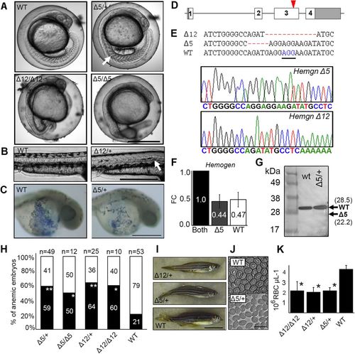
CRISPR/Cas9 mutagenesis of the third exon of zebrafish Hemogen reduces primitive and definitive erythropoiesis. Embryos were injected with Cas9 mRNA and a guide RNA to establish lines with mutations in exon three of zebrafish Hemogen. (A) 20 hpf. Representative wild-type and mutant siblings with notochord defects (arrow). (B) 48 hpf. Mutant Δ12 embryos with an in-frame deletion showing kinked notochords (arrow). (C) 24 hpf. Wild-type and Δ5/+ mutant embryos stained with diaminofluorene. Production of erythrocytes was reduced in heterozygotes. (D) Schematic of CRISPR/Cas9 target in the third exon (red arrowhead) of zebrafish Hemogen. (E) Sequences of founder mutations aligned at the CRISPR target site: Δ5 (Hemgnnuz2); Δ12 (Hemgnnuz4). The sequence traces show the Δ5 and Δ12 mutant alleles. PAM, blue and underlined; Δ, deletions (highlighted in red). (F) Relative expression of wild-type and Δ5 transcripts in blood from single adult, heterozygous Hemgnnuz2/+ mutants determined by qRT-PCR with allele specific primers. Three biological replicates were normalized to β-actin. Error bars represent the standard deviation. (G) Western blot of Hemogen in pooled 33 hpf wild-type embryos or pooled embryos from a Δ5 Hemgnnuz2/+ heterozygous in-cross. We calculated that the protein would run 6.5 kDa above its molecular weight at 28.5 kDa because of its high acidic composition (Guan et al., 2015). Arrows show the calculated sizes of wild-type and truncated alleles. (H) Proportion of genotyped mutants and wild-type sibling embryos at 2 dpf that were anemic (black) or phenotypically normal (white) (*P≤0.05, **P≤0.005, Chi square). (I) Wild-type and mutant zebrafish heterozygous for the Δ5 and Δ12 alleles. (J) Red blood cells from adult Hemgnnuz2/+ mutant zebrafish and wild-type siblings. (K) Erythrocyte counts in adult heterozygous Hemgnnuz2 (Δ5, n=12), heterozygous Hemgnnuz4 (Δ12, n=4) mutants, homozygous Hemgnnuz4 (Δ12, n=2) mutants and wild-type (n=9) siblings (*P≤0.05, ANOVA, Tukey post hoc test). Scale bars: 500 µm (A-C); 50 mm (I); 20 µm (J). |
| Fish: | |
|---|---|
| Observed In: | |
| Stage Range: | 20-25 somites to Adult |