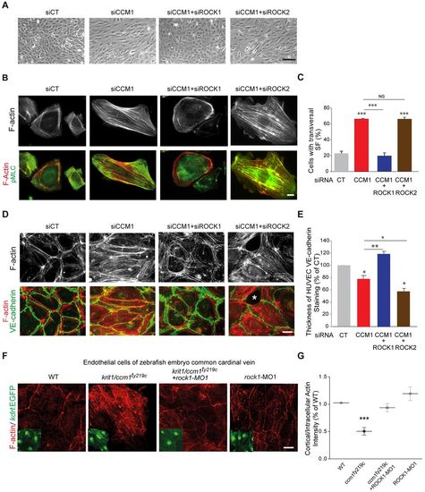
ROCK1 silencing restores normal adhesive and contractile phenotypes in CCM1-depleted HUVECs and zebrafish ccm1 mutant CCV endothelial cells. (A) Representative phase contrast images of siRNA-transfected HUVEC monolayers. (B) Representative immunofluorescence of sparsely plated siRNA-transfected HUVECs stained for F-actin (gray) and merged images with pMLC. (C) Percentage of cells with transversal actin stress fibers. Error bars, s.e.m. (n=4 except for CCM1+ROCK2 and CCM2+ROCK2 where n=2). (D) Representative immunofluorescence of F-actin (gray) and merged images with VE-cadherin of HUVEC monolayers on 20 μg/ml FN-coated coverslips at 72 h post plating. The asterisk indicates the zone of detachment for intercellular junctions. (E) Quantifications of VE-cadherin thickness from immunofluorescence staining of HUVECs presented as a percentage of control. The CT and CCM1 data are also shown in Fig. 1I. Error bars, s.e.m. (n=4 except for CCM1+ROCK1 where n=3 and CCM1+ROCK2 where n=2). In C and E, *P<0.05, **P<0.005, ***P<0.0005; NS, not significant (one-way ANOVA with Dunett's multiple comparisons). (F) Representative immunofluorescence of F-actin staining and Tg(kdrl:EGFP)-labeled endothelial cells (insets) of the CCV. (G) Quantification dot plot with s.e.m. of the intensity ratios of peripheral cortical actin over intercellular actin in WT (n=13), krit1/ccm1ty219c (16 embryos, 42 cells), krit1/ccm1ty219c+rock1-MO (16 embryos, 47 cells), and WT+rock1-MO (12 embryos, 26 cells) CCV regions; results were normalized to the WT ratio and the experiment was performed three times on independent zebrafish cohorts. ***P<0.005 (one-way ANOVA with Tukey's multiple comparisons post-test). Scale bars: 50 μm (A) 10 μm (B,D,F). CT, control.
|
