
FIGURE
Fig. 6
- ID
- ZDB-FIG-180917-29
- Publication
- Svahn et al., 2018 - Nucleo-cytoplasmic transport of TDP-43 studied in real time: impaired microglia function leads to axonal spreading of TDP-43 in degenerating motor neurons
- Other Figures
- All Figure Page
- Back to All Figure Page
Fig. 6
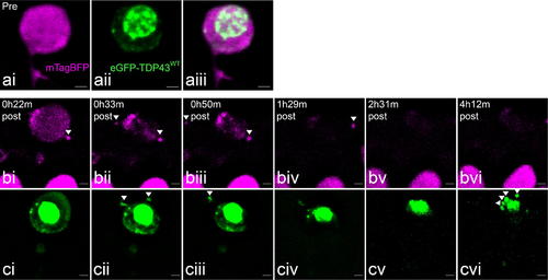
Fragment release from a neuron undergoing UV-induced degeneration in the absence of functional microglia. ai Cytoplasmic mTagBFP signal pre-irradiation. aii eGFP-TDP43WT distribution in the soma pre-irradiation. aiii Merged images. bi–vi mTagBFP during UV-induced degeneration. Concentrated fragments can be observed separating from the external membrane before the cytoplasm disperses. ci–vi eGFP-TDP43WT during degeneration. Concentrated fragments can be observed collecting and releasing from the membrane. The concentrated (pyknotic) nuclear core gradually fragments. Scale = 2 µm |
Expression Data
Expression Detail
Antibody Labeling
Phenotype Data
Phenotype Detail
Acknowledgments
This image is the copyrighted work of the attributed author or publisher, and
ZFIN has permission only to display this image to its users.
Additional permissions should be obtained from the applicable author or publisher of the image.
Full text @ Acta Neuropathol.