
FIGURE
Fig. 4
- ID
- ZDB-FIG-180913-2
- Publication
- Nadarajah et al., 2018 - A Novel Splice-Site Mutation in VEGFC Is Associated with Congenital Primary Lymphoedema of Gordon.
- Other Figures
- All Figure Page
- Back to All Figure Page
Fig. 4
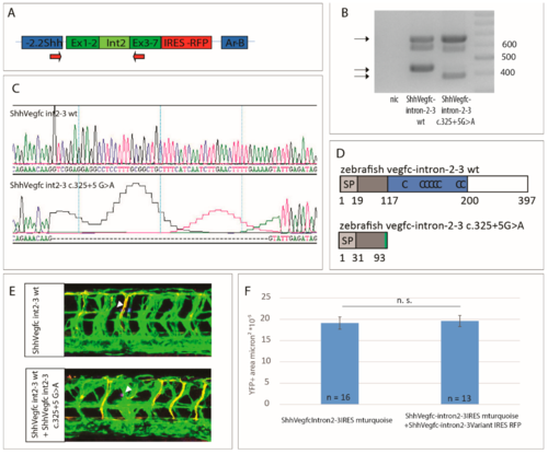
The vegfc c.325+5G>A splice-site variant results in a truncated protein, which does not have dominant negative activity. (A) Diagram of the construct used for overexpression of vegfc-intron 2–3 depicting primers used for RT-PCR (arrows). (B) RT-PCR of zebrafish embryos expressing ShhVegfc-intron 2–3 wt or ShhVegfc-intron 2–3 c.325+5G>A. Embryos expressing the wt form of ShhVegfc-intron 2–3 and the ShhVegfc-intron 2–3 c.325+5G>A variant express a band of 660 bp corresponding to non-spliced RNA or integrated plasmid DNA as well as a second smaller band corresponding to wt ShhVegfc (440 bp) or mutant ShhVegfc (387 bp). Right lane: 100 bp ladder. Non-injected control, nic. (C) Sequencing of cDNA of embryos expressing ShhVegfc-intron 2–3 and the ShhVegfc-intron 2–3 c.325+5G>A variant showing a 53 bp deletion in the ShhVegfc-intron 2–3 c.325+5G>A variant (lower panel). (D) Schematic representation of predicted wild type (top) and mutant (bottom) proteins. Mutant protein consists of the first 91 amino acids of Vegfc containing only a part of the N-terminus of Vegfc but not the VHD or the C-terminus (see Figure 2 for legend). (E) Analysis of co-overexpression of ShhVegfc-intron 2–3 wt and ShhVegfc-intron2–3 c.325+5G>A in the floorplate using the transgenic line Tg(flt4BAC:mCitrine)hu7135 and Tg(flt1enh:tdTomato), which marks venous and lymphatic cells in green and arterial vessels in red at 48 hpf. Expression of ShhVegfc-intron 2–3 wt and ShhVegfc-intron 2–3 c.325+5G>A within the same cell are monitored by the simultaneous expression of mturquoise and tagRFP, respectively. Forced expression of ShhVegfc-intron 2–3 wt in the floorplate led to excessive vessel sprouting comparable with co-overexpression of ShhVegfc-intron 2–3 wt and ShhVegfc-intron 2–3 c.325+5G>A in the floorplate. Arrow heads mark expression of tagRFP and mturquoise in the floorplate. (F) Quantification of lymphovenous sprouting by measuring the YFP positive area surrounding the site of mturquoise/tagRFP expression. Values are presented as means ± standard error of mean values (SEM). n.s. not significant.
|
Expression Data
Expression Detail
Antibody Labeling
Phenotype Data
Phenotype Detail
Acknowledgments
This image is the copyrighted work of the attributed author or publisher, and
ZFIN has permission only to display this image to its users.
Additional permissions should be obtained from the applicable author or publisher of the image.
Full text @ Int. J. Mol. Sci.